Description
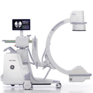
The GE OEC 9800 C-Arm system offers a high resolution screen, touch-screen controls, multiple configurations, physical options and increased storage. This advanced system enhances medical imaging with real-time anatomy tracking, automatic collimator position detection, and adaptive metal enhancement. It features precise tungsten collimation for X-ray control and safety in Orthopedic, General, Pain Management, Spine, and Peripheral Vascular procedures. The system boasts a high-resolution 1k x 1k camera with motorized rotation and user-friendly touchscreen controls.
X-Ray System:
Generator:
60kHz high frequency
15kWpower
Up to 120kVp
Up to 75mA for radiographic film exposure
Continuous high-level fluoro (HLF) up to 20mA
Digital spot up to 75mA
Digital cine pulse:
15 pulses per second, 60 Hz
12 pulses per second, 50 Hz
Up to 150mA
10ms pulse width
Full power from standard wall outlet
Patented Battery buffered design
X-Ray Tube:
Rotating anode x-ray tube
0.3 and 0.6 nominal focal spots
Anode heat capacity: 300,000 HU
Anode cooling rate: 85,000 HU/min
Housing heat capacity: 1,600,000 HU
9″ (23cm) Standard C-Arm housing cooling rate 15,000 HU/min
9″ (23cm) Super C-Arm and 12″ (31cm) Standard C-Arm housing cooling rate 22,500 HU/min
Digital Image Rotation:
Digitally adjusts image display
Image update
Image rotation
Image reversal (side-to-side)
Image invert (top-to-bottom)
Image positioning without additional exposure
PreView Collimator:
On-screen collimator position indication
PreView iris collimator
PreView Tungsten rotatable double leaf collimator
Adjusts collimators without x-ray exposure
Video Imaging System:
9″ Image Intensifier:
Tri-mode 9″/6″/4.5″ (23cm/15cm/11cm) image intensifier
Minimum central resolution (at the monitor):
9″ (23cm): 2.1 lp/mm
6″ (12cm): 2.9 lp/mm
4.5″ (11cm): 3.4 lp/mm
DQE: 65% (typical)
12″ Image Intensifier:
Tri-mode 12″/9″/6″ (31cm/23cm/15cm) image intensifier
Minimum central resolution (at the monitor):
12″ (31cm): 1.5 lp/mm
9″ (23cm): 2.1 lp/mm
6″ (12cm): 2.9 lp/mm
DQE: 65% (typical)
Autotrak Automatic Brightness Stabilization (ABS):
Automatically seeks the subject anatomy anywhere within the imaging field and selects the optimum imaging technique
Automatically adjust to anatomical size and location
Provides uniform image quality throughout entire image
Simplifies operation
Image I.Q.:
Smart Window:
Dynamically senses the collimator position and automatically adjusts brightness and contrast to produce high image quality.
Smart Metal:
Allows user to adjust automatic brightness and contrast sensitivity levels to metal
Provides optimum image quality even when metal is introduced to the field
Tungsten Collimator:
Denser collimator limits x-ray exposure area
Reduces scatter radiation
Improves image detail
Video Camera:
High resolution 1k x 1k CCD camera
Full frame capture
Motorized rotation
On-screen orientation indicator (real-time feedback without fluoro)
Left-right image reversal
Top-bottom image invert
Video Display (OEM Factory):
Dual 16″ (41cm) square monitors
Anti-glare, progressive full frame scan monitor
Touch screen system control
1,000 line high resolution monitors
Ambient room-light compensation
Video Display (Upgraded Retrofit Kit):
32″ LED Lit LCD Display with Dual Screen Capabilities
Anti-glare, progressive full frame scan monitor
Touch screen system control
2 Mega Pixel high-resolution output
Ambient room-light compensation
Modes of Operation Fluoro Mode:
kVp range: 40-120
mA range:
0.2-10 normal mode
0.2-20 HLF (high level fluoro)
Auto and manual fluoro modes
AutoTrak ABS varies mA, kVp, camera gain
Pulsed Fluoro Mode:
kVp range: 40-120
mA range: 0.2 -10
Pulse rate: 1, 2, 3, 8
Pulse width: 25 or 50 ms
AutoTrak ABS, mA, kVp, camera gain
Reduces x-ray dose to patient and operator
High Level Pulsed Fluoro:
kVp range: 40-120
mA range: 0.2-40
Pulse rate: 1, 2, 4, 8
AutoTrak ABS, mA kVp, and camera gain
Digital Cine Pulse Mode:
kVp range: 40-120
mA range: up to 150
Pulse rate: 15 pps (60 Hz), 12 pps (50Hz)
Pulse width: 10ms
AutoTrak ABS, mA, kVp, camera gain
Digital Spot Mode:
kVp Range: 40-120
mA range: up to 75
Automatic exposure termination and automatic image save
Radiographic Mode:
mA range: up to 75
mAs range: up to 300
Computer controlled exposure time
Optional film casette holder
10×12″ for 9″ II
14×14 for 12″ II
Configurations GSP (General Surgical Package) Platform:
1k x 1k x 16 bit image processing
Noise filter with on-screen indicator
Real-time variable edge enhancement
Automatic digital brightness and contrast control
Negate mode
Save and auto-save feature
Swap and auto-swap feature
Patient information
Examination list
Customized patient information
Customize functions:
Workstation set-up
Mainframe set-up
Patient information set-up
Date/time set-up
DICOM 3.0 interface set-up
Last image hold
63 image storage
Integrated DICOM interface (store, print, worklist)
HIPAA SecureView:
Password protection
Blank screen function
Delete all
ESP (Expanded Surgical Package) Platform:
Includes all the GSP features and:
200 or 400 image storage
Zoom and roam function
Image annotation
Measure software
PMCare Platform:
Includes all the ESP features and:
Real-time digital subtraction (DSA)
Peak opacification
8fps dynamic digital disk
Recording /playback rate: 1, 2, 4, 8fps
Recording time: 5 minutes @ 8fps
Vascular Platform:
Includes all the ESP features and:
Real-time digital subtraction (DSA)
Roadmapping
Peak opacification
Re-registration
Variable landmarking
Mask save/recall
Auto cine loop playback
Digital cine pulse mode:
15 pulses/sec
Up to 150mA
10ms pulse width
15fps dynamic digital disk:
Recording/playback rate: 1, 2, 4, 8, 15fps
Recording time: 10 minutes @ 15fps (time depends on record frame rate)
Neuro-Vascular Platform:
Includes all the Vascular features and:
30fps dynamic digital disk:
Recording/playback rate: 1, 2, 4, 8, 15, 30fps
Recording time: 10 minutes @ 30fps (time depends on record frame rate)
Cardiac Platform:
Includes all the Neuro-Vascular features and:
Single leaf curved collimator
Three pedal footswitch
Upgraded x-ray tube cooling
Additional Features Additional Features:
Super-C Configuration:
Adds Larger “C” for increased orbital rotation (145 degrees)
Increased Arc Depth (33″ / 838mm)
MD Configuration:
Adds motorized movements of c-arm
Other Image Storage Options:
CD/DVD Writer digital interface (external)
Integrated DICOM 3.0 interface
Hardcopy Options:
Integrated film/paper printer
-No film developing required
-8×10″ (20.3cm x 25.5 cm) laser quality film/paper
-Multi-format, 1,2, 4 on 1
-Multi-copy capability
Thermal printers
User Interface:
Entire system is computer controlled and sofware upgradeable
Touchscreen control simplifies operation
Automated system operation requires minimum operator interface
Multi-functional controls
Footswitch
IR remote (optional)
Hand-held control
Simplified keyboard
Multi-Purpose image directory
Retrieve and review images
Compose hardcopy films
Copy images
X-ray dose summary
Electrical:
Input power (69Hz ir 50Hz)
100v @ 20 A (Japan)
120V @ 12 A
200V, 220V, 230V, 240V @ 10 A
Regulatory Compliance:
U.S. 21 CFR Subchapter K
NFPA 99
UL 60601-A (CSA/NRTL)
IEC60601-1 (plus relevant collateral and particular standards)